A New Brain Aneurysm Cell Atlas Offers Clues About Why Some Blood Vessels Become Vulnerable
Researchers have created a detailed cellular map of brain aneurysm tissue, offering new insight into how certain blood vessel walls may become fragile over time.
Save Article
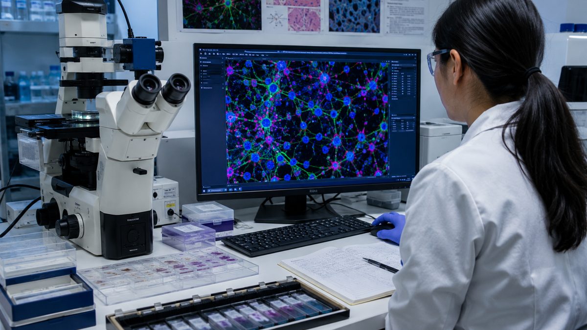
Cell-level research can help scientists study how fragile blood vessels change over time. Editorial illustration by TheDailyGlobe.
Key Facts
- The study was published in Nature Neuroscience on June 10, 2026.
- Researchers used single-cell and spatial transcriptomics to analyze aneurysm tissue.
- The work examined cellular activity within human brain aneurysms.
- Researchers identified links between scarring-related fibroblasts and inflammatory macrophages.
- The findings focus on disease mechanisms and do not represent a new diagnostic test or treatment.
A weak spot in a blood vessel can exist for years without causing symptoms. In some cases, that weakness develops into a brain aneurysm, a condition that can remain hidden until it is discovered during imaging or, in more serious situations, after a rupture.
Scientists have long known that aneurysms involve changes in the walls of blood vessels. What has been harder to understand is exactly what is happening inside those tissues at the cellular level. A new study published in Nature Neuroscience offers one of the most detailed looks yet at that question.
Using advanced mapping techniques, researchers created a cellular atlas of human brain aneurysm tissue, providing a closer view of the cells that may contribute to vessel wall changes over time.
A Closer Look Inside Vulnerable Vessel Walls
The central goal of the study was not to test a treatment or evaluate patient outcomes. Instead, researchers wanted to better understand the biological environment inside aneurysm tissue.
To do that, the team analyzed tissue at an unusually detailed level. Rather than treating a blood vessel wall as a single structure, they examined the different cell populations present within it and how those cells interacted with one another.
The resulting atlas allowed researchers to identify patterns that may help explain how vessel walls are remodeled as disease progresses. Among the findings was evidence linking scarring-related fibroblasts and inflammatory macrophages to changes within aneurysm tissue.
How Scientists Built the Atlas
The study relied on two technologies that have become increasingly important in modern biomedical research: single-cell transcriptomics and spatial transcriptomics.
In simple terms, these tools help scientists examine which genes are active within individual cells while also showing where those cells are located within a tissue sample. Instead of seeing only a broad average across an entire specimen, researchers can observe distinct cell types and how they are arranged.
That combination provides a more detailed picture of biological activity. It allows scientists to study not only which cells are present, but also how neighboring cells may influence one another inside diseased tissue.
Why the Findings Matter
Understanding disease mechanisms is often one of the earliest steps in medical research. Before researchers can develop better prediction tools, therapies, or preventive strategies, they first need to understand what is happening inside affected tissues.
The new atlas contributes to that effort by providing a more detailed map of the cellular environment associated with brain aneurysms. Researchers hope such information may eventually help identify biological pathways that deserve further study.
For readers, the value of the study is not that it changes medical care today. Rather, it offers insight into how scientists investigate diseases that can be difficult to observe directly.
What the Research Does Not Prove
The study comes with important limitations. It does not provide a new screening test, treatment recommendation, or prevention strategy for patients.
Researchers identified cellular patterns associated with vessel wall remodeling, but translating those findings into practical medical tools could take years of additional research. The study was designed to improve understanding of biological mechanisms, not to establish clinical guidance.
Scientists also caution that promising laboratory findings do not automatically become useful medical interventions. Many discoveries help advance understanding without ultimately leading to new treatments.
The Questions Researchers Are Still Asking
Several important questions remain unanswered. Researchers do not yet know how quickly the findings could influence future clinical tools, if at all. It is also unclear whether additional studies will confirm the same cellular patterns across broader patient populations.
Future research will likely focus on validating the findings, exploring the biological pathways involved, and determining whether any of the identified cellular signals could eventually serve as biomarkers or therapeutic targets.
For now, the study represents a deeper look into a disease process that scientists are still working to understand. The most important advance is not a new treatment, but a clearer map of the cellular landscape researchers are trying to navigate.
Reporting note: Reporting draws on peer-reviewed research, scientific journal materials, biomedical reporting, and reviewed background materials. This article was produced with AI-assisted research and reviewed by an editor before publication.